Need ideas of where to read up on the latest medical education research? Have a brilliant manuscript describing your curricular innovations but not sure where to publish? Need to find multimedia resources for medical education? Looking for curricular objectives to help guide your medical education project?
Look no further than the Medical Education Subject Guide available on the UCSF Library's website. The direct URL to this guide is at http://guides.library.ucsf.edu/meded.
OR you can navigate from the Library's website from the Classes, Consulting, & Help dropdown menu by selecting the Subject Guides listing. The Medical Education guide is listed alphabetically on the complete guides page.
(1) Browse the yellow tabs to explore the Library's resources available to assist in your medical education research needs.
(2) Read the latest published medical education research by UCSF medical educators.
(3) Download PDF tipsheets summarizing literature searching tips for how to search PubMed and other key databases geared to medical education.
(4) Contact me via chat, email, or phone from the right margin of the guide.
Please feel free to send me any suggestions you have for content you'd like to see on this subject guide. Bookmark this site today! http://guides.library.ucsf.edu/meded
Showing posts with label images. Show all posts
Showing posts with label images. Show all posts
Wednesday, July 6, 2011


Thursday, March 25, 2010
iRadiology App

The iRadiology App is available for free to download at the iTunes Apps store.
This app allows you to browse amongst 500+ radiology cases to improve skills to interpret plain film, CT, and MRI readings.
The cases are based upon materials from Dr. Gillian Lieberman, the Director of Harvard Medical Student training and Associate Director of the Residency Program at Beth Israel Medical Center.
You can increase the zoom capability on your iPhone to better view the images:
- Go to "Settings", tap "General", then "Accessibility"
- Tap "Zoom", and slide the Zoom feature to the "On" position

Wednesday, October 28, 2009
Free Mind Mapping Tools
Oh boy - I've found some scrumptious free mindmapping tools that I need to share with you!
Mind mapping is a way to visualize concepts by drawing out associations between your ideas. It can be extremely useful as a brainstorming tool and even for teaching. It combines the textual and visual cognitive senses we use to help us understand concepts.
Two absolutely free and absolutely easy to use mind mapping tools are:
(1) bubbl.us - you don't need to set up an account. Simply click on the "Start Brainstorming"button.
The controls are intuitive. For example, hovering over the first box allows you to type in text. You can hover over parts of that box to show crosshairs that allow you to reposition the box wherever you want. The other hovering options allow you to add boxes stemming from the original. You can change the color of the boxes.
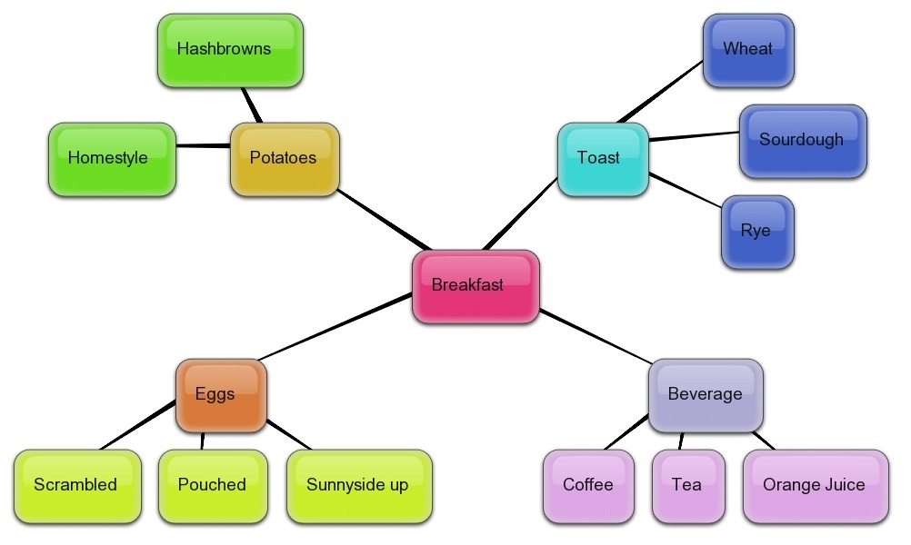
When you're done mapping, click on the Menu button at the bottom right of the screen. You can export the map you've created as an image, XML, or HTML file. Below is an example of a mind map I created using bubbl.us:
(2) SimpleMind Xpress - this is a free app for your iPhone or iPod Touch. There is a paid version, but I find the free one does a good job. You can upload your final mind map to your iTunes and then download it to your computer as a document.

This presentation below gives some helpful pointers on how to maximize the effectiveness of your mind mapping exercises.
Mind Mapping
View more presentations from Amir Saif .
Tuesday, September 29, 2009
Google Quick Tip #23
Behold - there's a way to sort through the data dump you get when
you run a Google Images search!
GOOGLE QUICK TIP #23
The next time you run a Google Images search, click the Show Options link on the left above your list of results.

A list of options will appear in the left menu. You can filter the image results to your own specifications, such as size, color, and image type.
Click on the Reset options link at the bottom of the left menu if you want to return to your original set of image search results. And last but not least, remember to always site your image sources!
View this quick narrated tutorial to see this feature in action.
Monday, September 28, 2009
EdHeads Virtual Surgery Games
If you're not a real surgeon, but you want to play one (on the web), EdHeads.org has created a site that's just for you.

The EdHeads team creates educational interactive web modules. There are currently nine activities to choose from. The medically related ones are surgical deep brain stimulation, hip replacement, hip resurfacing, and knee surgery.
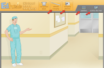
Once you've entered one of the surgical activities, you'll be taken from the Reception Desk into the hallway of the OR. A virtual surgeon will orient you to your patient and have questions for you to interact with and answer throughout the tutorial. You will be asked to select different surgical instruments and use them during the surgery.
The tutorial is narrated, but you can also select the Subtitles icon to see closed captioning. If you want to skip ahead to different parts of the tutorial, select the Activity Site Map icon.
If you have the stomach to view actual photos of the surgeries being described, click on the Surgery Photos icon.
Included with each activity is a Teacher's Guide to give you ideas of how to make the activities a useful learning tool for your students. There are sample quiz questions and answers, a glossary, and additional helpful resources on the topic.
If you're in the mood for something non-medical, check out the others, such as Designing a Cell Phone or learning how to predict and report the Weather.
Be inspired to have fun!
Thursday, July 30, 2009
Clinical Cases and Images blog
The Clinical Cases and Images blog is a "free case-based curriculum of clinical medicine." I stumbled upon this resource three years ago and have recommended it as a reference to students for finding clinical images that are accompanied by helpful discussions.

The blog was created in 2005 and is a joint effort of physicians at the Cleveland Clinic and the Case Western Reserve School of Medicine. It is well organized by specialties along the top menu bar.
The topics covered include:
Allergy & Immunology -- Cardiology -- Pulmonology -- Gastroenterology -- Nephrology -- Endocrinology -- Hematology & Oncology -- Rheumatology -- Infectious Diseases -- Neuorology -- Geriatrics -- Pain Management
Other categories offered provide more information and links in the right menu bar:
-- Physical Examination Videos
-- Procedure Guides Step-by-Step
-- Electrocardiograms, X-rays, CT scans
-- Clinical Images from NEJM
-- Clinical Cases in Test Format (test your knowledge based on details of a case)
In the lower part of the right menu bar is a link to many of the images found in the blog that are housed on the photo hosting site Flickr - http://www.flickr.com/photos/clinicalcases/.
The comments made on the content are helpful discussions. An encouraging feature to note is that all entries have a "Published" and "Last Updated" dates that you'll find by scrolling down the page. This can help you evaluate the usefulness and timeliness of the content.
To get a sampling of what you can learn from this well done blog, take a look at EKGs with Dr. Koch: It's Not Only Educational It's Also Fun!
Monday, June 29, 2009
DermAtlas
Dermatology image resources may prove hard to find in the vast web of online information. Here's one that I came across recently that looks very well organized and informative.
 DermAtlas is a Dermatology Image Atlas created by Dr. B.A. Cohen and Dr. C.U. Lehmann of Johns Hopkins University. The site boasts over 11,000 dermatology images from 513 physicians and other health professionals from 75 nations.
The basic Quick Search box is a good place to start if you know exactly what you're looking for. I recommend trying out some of the features in the left menu.
- Adv.Search : can limit your search by several criteria, such as body site, pigmentation, age, gender, and histology.
- New Images : lists images submitted in the last few months
- Differential : this search tool claims to help you figure out what type of rash you see. It is still under construction, so they provide a caveat that you search at your own caution. You can limit by morphology, color, pattern, organization, and location.
The best way to find out about an image is to click on the image itself to get detailed information. Some of the detailed information is hyperlinked and takes you to other images related to that topic.
Very helpful resource links are located at the bottom in the Other Resources portion. There you'll find links to more information about the condition in PubMed, MedlinePlus, eMedicine, Archives of Dermatology, Dermatology Online, and OMIM (the genetics database component related to PubMed).
There is an option to translate the image description near the very bottom of the page. Languages include German, Spanish, Italian, French, and Portuguese.
At the very bottom of the page are related images that you can click on and learn more. In the left menu, you'll also find links to quizzes and CME credits for continuing education.
I'm no expert on dermatology, so if you check out this resource, let me know how it ranks in your opinion. Feel free to leave a comment on your own recommendations!
DermAtlas is a Dermatology Image Atlas created by Dr. B.A. Cohen and Dr. C.U. Lehmann of Johns Hopkins University. The site boasts over 11,000 dermatology images from 513 physicians and other health professionals from 75 nations.The basic Quick Search box is a good place to start if you know exactly what you're looking for. I recommend trying out some of the features in the left menu.
- Adv.Search : can limit your search by several criteria, such as body site, pigmentation, age, gender, and histology.
- New Images : lists images submitted in the last few months
- Differential : this search tool claims to help you figure out what type of rash you see. It is still under construction, so they provide a caveat that you search at your own caution. You can limit by morphology, color, pattern, organization, and location.
The best way to find out about an image is to click on the image itself to get detailed information. Some of the detailed information is hyperlinked and takes you to other images related to that topic.
Very helpful resource links are located at the bottom in the Other Resources portion. There you'll find links to more information about the condition in PubMed, MedlinePlus, eMedicine, Archives of Dermatology, Dermatology Online, and OMIM (the genetics database component related to PubMed).
There is an option to translate the image description near the very bottom of the page. Languages include German, Spanish, Italian, French, and Portuguese.
At the very bottom of the page are related images that you can click on and learn more. In the left menu, you'll also find links to quizzes and CME credits for continuing education.
I'm no expert on dermatology, so if you check out this resource, let me know how it ranks in your opinion. Feel free to leave a comment on your own recommendations!
Friday, May 29, 2009
Yale Image Finder
Gosh - I love technology! Think of an idea, and most likely someone's come up with an answer. Or at least a pretty awesome prototype.
YALE IMAGE FINDER is a search tool that allows you to look for images and figures from free full-text articles in the PubMed Central database of Open Access articles.
This tool debuted in July 2008 as a project of the Krauthammer Lab of the Yale Pathology Informatics Department that specializes in text mining and translational informatics.
View this short narrated tutorial on how to search Yale Image Finder:

The Krauthammer Lab published a paper that describes their search algorithm:
ABSTRACT: Yale Image Finder (YIF) is a publicly accessible search engine featuring a new way of retrieving biomedical images and associated papers based on the text carried inside the images. Image queries can also be issued against the image caption, as well as words in the associated paper abstract and title. A typical search scenario using YIF is as follows: a user provides few search keywords and the most relevant images are returned and presented in the form of thumbnails. Users can click on the image of interest to retrieve the high resolution image. In addition, the search engine will provide two types of related images: those that appear in the same paper, and those from other papers with similar image content. Retrieved images link back to their source papers, allowing users to find related papers starting with an image of interest. Currently, YIF has indexed over 140,000 images from over 34,000 open access biomedical journal papers.
URL: http://krauthammerlab.med.yale.edu/imagefinder/
Wednesday, April 29, 2009
Bassett Collection (Human Anatomy)
Have a strong stomach for detailed anatomy images? The Bassett Collection of Stereoscopic Images of Human Anatomy is now available on the Stanford Lane Medical Library's website.
This is an astounding collection of 1,547 photographs with accompanying sketches and descriptions. Information about the collection:
"David Lee Bassett (1913-1966) was awarded his MD by Stanford University School of Medicine in 1939. He began teaching in the Department of Anatomy at Stanford University School of Medicine in 1939 and later also taught anatomy at the University of Washington in Seattle. In 1948 Bassett met William B. Gruber, inventor of the Viewmaster system of stereoscopic imagery. Bassett and Gruber collaborated on a seventeen-year project of creating three-dimensional photographic images of human anatomy using innovations in dissection pioneered by Dr. Bassett."
Any questions about permissions of use should be directed to Drew Bourn at dbourn@stanford.edu.
This is an astounding collection of 1,547 photographs with accompanying sketches and descriptions. Information about the collection:
"David Lee Bassett (1913-1966) was awarded his MD by Stanford University School of Medicine in 1939. He began teaching in the Department of Anatomy at Stanford University School of Medicine in 1939 and later also taught anatomy at the University of Washington in Seattle. In 1948 Bassett met William B. Gruber, inventor of the Viewmaster system of stereoscopic imagery. Bassett and Gruber collaborated on a seventeen-year project of creating three-dimensional photographic images of human anatomy using innovations in dissection pioneered by Dr. Bassett."
Any questions about permissions of use should be directed to Drew Bourn at dbourn@stanford.edu.
Wednesday, March 25, 2009
Turning the Pages of Historical Medical Texts
At the National Library of Medicine in Washington D.C. there is an electronic kiosk that allows visitors to browse the pages of historical medical textbooks. This Turning the Pages project is available now online for you to browse on your computer.
The interactive books are quite beautiful. For example, you can flip through the pages of Andreas Vesalius's De Humani Corporis Fabrica (On the Fabric of the Human Body). This book is revered as "one of the most influential books in the history of Western medicine."
'It was conceived and written by 28-year-old Andreas Vesalius (1514-1564), a professor at the University of Padua. Vesalius was both a gifted dissector and a learned scholar whose great contribution was to apply to anatomy the critical methods developed by the Renaissance humanist scholars."
Below is one of the famous images from Vesalius's series of skeletal men. This one shows a skeleton "contemplating human mortality."
The interactive books are quite beautiful. For example, you can flip through the pages of Andreas Vesalius's De Humani Corporis Fabrica (On the Fabric of the Human Body). This book is revered as "one of the most influential books in the history of Western medicine."
'It was conceived and written by 28-year-old Andreas Vesalius (1514-1564), a professor at the University of Padua. Vesalius was both a gifted dissector and a learned scholar whose great contribution was to apply to anatomy the critical methods developed by the Renaissance humanist scholars."
Below is one of the famous images from Vesalius's series of skeletal men. This one shows a skeleton "contemplating human mortality."

- Click on a page to advance to the next page. Click on the left page to go back a page.
- Unless you can read Latin, I suggest you click on the speaker icon to hear the narrator describe content on the pages you are viewing.
- Use the magnifying glass icon to zoom over the text.
- Hover the mouse over sections of the page and you can read curatorial notes describing the content.
- Click the "T" icon to the left of the page to see the transcript of the audio for that page.
- You can also print a page if you wish.
Even if you're not a historical book fanatic, I encourage you to browse these pages. It's a privilege to be able to see the details of a book written in 1543.
Saturday, February 28, 2009
Google Quick Tip #17
How many ways can you slice your search results? Well, Google can show you by displaying your results in different formats.
GOOGLE QUICK TIP #17
By typing in the phrase view:timeline after your search words, your results will display as seen below.
For example, I searched for harold varmus view:timeline
You can also select any of the other options above the graph to display your results in a list, info, or map view.
For you history buffs, check out the results for a search on world war 2 view:timeline
GOOGLE QUICK TIP #17
By typing in the phrase view:timeline after your search words, your results will display as seen below.
For example, I searched for harold varmus view:timeline

* The graph summarizes how dates in your results are spread through time
* The higher bars represent a larger number of unique dates
* Click time periods on the graph to zoom in to that particular period of time
* Can search the text box to the right to specify any range of years, months, or days
You can also select any of the other options above the graph to display your results in a list, info, or map view.
For you history buffs, check out the results for a search on world war 2 view:timeline
Learn Anatomy with WinkingSkull.com
Looking for a good learning tool to help you learn and test yourself on human anatomy? The WinkingSkull.com offers just that!

This free resource has detailed color images from the Atlas of Anatomy. When you create a free account, you get the full capability to test yourself on what you've learned.
(1) Go to the "My Account" link at the top right to create a free account
(2) Select the region of the body that you're interested in viewing via the left menu
(3) Select the labels to be "ON"
(4) You can also view the labels in Latin by selecting the "US English" box at the top right and switch to "Latin"
(5) You can zoom in and out, as well as hold down the mouse and move around the picture
(6) When you are signed into your account, you can test your knowledge of the anatomy you've reviewed
Be sure to check out their Quick Tips link in the top menu bar.
Watch this short non-narrated video to view these features [click on the "full size" link].

Monday, June 30, 2008
Radiology Picture of the Day (blog)
Like most internet users, you probably grab images from a basic Google image search. Well, thank your lucky stars that there are some nice alternatives to just "googling" for medical images.
Check out the Radiology Picture of the Day blog. A new image is posted daily that is provided by a physician with a brief description below about the image with related references.
The blog is edited and maintained by Dr. Laughlin Dawes, a diagnostic neuroradiology fellow at the Sir Charles Gairdner Hospital in Perth, Western Australia. The images date back from November 2006 to the present.
I prefer to navigate this blog via its ARCHIVE page that categorizes the images for you. There is a link to suggest an image that you would like to submit to this blog, as well.
As always, give credit where it's due if you are going to use any image from the web. For example, if I were to use this image

I would reference this image as:
Congenital cystic adenomatoid malformation. Image provided by Dr. Ahmed Haroun. Posted Feb 28, 2008 on the Radiology Picture of the Day blog. Accessed on June 30, 2008 at http://www.radpod.org/2008/02/28/congenital-cystic-adenomatoid-malformation.
Check out the Radiology Picture of the Day blog. A new image is posted daily that is provided by a physician with a brief description below about the image with related references.
The blog is edited and maintained by Dr. Laughlin Dawes, a diagnostic neuroradiology fellow at the Sir Charles Gairdner Hospital in Perth, Western Australia. The images date back from November 2006 to the present.
I prefer to navigate this blog via its ARCHIVE page that categorizes the images for you. There is a link to suggest an image that you would like to submit to this blog, as well.
As always, give credit where it's due if you are going to use any image from the web. For example, if I were to use this image

I would reference this image as:
Congenital cystic adenomatoid malformation. Image provided by Dr. Ahmed Haroun. Posted Feb 28, 2008 on the Radiology Picture of the Day blog. Accessed on June 30, 2008 at http://www.radpod.org/2008/02/28/congenital-cystic-adenomatoid-malformation.
Friday, May 30, 2008
Heart Sounds Tutorial from Blaufuss
THE HEART - so vital to our existance, so profound, so...hard to remember all the boatloads of information related to the heart! Well, thank goodness for tutorials such as the ones created by Blaufuss Multimedia Laboratories.

Their Heart Sounds and Cardiac Arrythmias tutorials walks you through all you need to know about heart sounds and electrocardiogram / arrhythmias. There are pictures, animated images, sound, and you can even take a quiz to test your knowledge.
Bless their hearts for creating such an awesome resource!
Subscribe to:
Posts (Atom)